Difference between revisions of "Lobule VIIAt"
Jump to navigation
Jump to search
(add category) |
|||
| (One intermediate revision by one other user not shown) | |||
| Line 1: | Line 1: | ||
| − | <meta name="title" content="Lobule VIIAt (Crus II)"/> | + | <!-- <meta name="title" content="Lobule VIIAt (Crus II)"/> --> |
{{cer nav|lobule=true}} | {{cer nav|lobule=true}} | ||
{{h2|Lobule VIIAt (Crus II)}} | {{h2|Lobule VIIAt (Crus II)}} | ||
Latest revision as of 00:58, 3 July 2022
Lobule VIIAt (Crus II)
- Location: Spans entire cerebellum about middle of the sagittal orientation
- Description: This lobule can first be identified and painted in the midsagittal and then the paint can be propagated laterally, can also be seen in the posterior axial slices.
- In the midsagittal region, there is an easy to define fissure separating this lobule and Lobule VIIIA.
- This fissure is primarily defined by the most dominate fissure between VIIAf and the prebiventure fissure, however determining which fissure qualifies can be difficult. Be patient and consider all perspectives and relavent regions of the scan in order to make the best decision possible.
- The best palace to discern this boundary is in the final figure below.


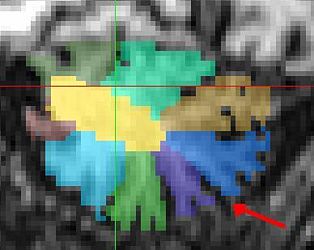
Figure 1:In my experience this is the best place to dicern the border between cruise 2 and VIIB
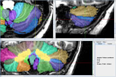
Figure 2:In this scan on the left 7B curves around Cr2 in a somewhat unusual manner.
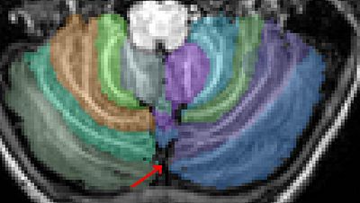
Figure 3 : The boundary between Cr2 and VIIB in the left hemisphere is particularilly difficult in this scan because there are branches crossing all possible boundaries.


