FANTASM: Fuzzy and Noise Tolerant Adaptive Segmentation Method
Dzung L. Pham and Jerry L. Prince
|
Overview | ||||||||||||||||||||||||||
| ||||||||||||||||||||||||||
|
Introduction | ||||||||||||||||||||||||||
|
Tissue classification is a necessary step in many medical imaging applications including the quantification of tissue volumes, the detection of pathology, and computer integrated surgery. Classification of voxels exclusively into distinct classes, however, is difficult because of artifacts such as noise and partial volume effects, which occur when multiple tissues contribute to a single voxel. To compensate for these artifacts, there has recently been growing interest in soft segmentation methods. In soft segmentations, voxels may be classified into multiple classes with a varying degree of membership. The Fuzzy C-Means clustering algorithm (FCM) is a soft segmentation method that has been used extensively for segmentation of magnetic resonance (MR) images because it is automatic and is applicable to a wide variety of problems. Standard FCM, however, can not effectively compensate for intensity inhomogeneities, a common artifact in MR images. Pham and Prince [1,2] proposed an algorithm called the Adaptive Fuzzy C-Means Algorithm (AFCM) that produces a soft segmentation while simultaneously adapting to intensity inhomogeneities in the image. The algorithm was derived by incorporating a gain field term into the objective function of the standard fuzzy c-means algorithm. Constraints on the gain field were used to ensure that the estimated field was smooth and slowly varying. While AFCM has been shown to be effective in correcting for inhomogeneities, its main disadvantage is that its performance degrades significantly with increased noise. One approach to solving this problem would be to preprocess the image with a smoothing filter. However, typical smoothing filters cause a loss of detail in the original image and are not mathematically consistent with the fuzzy clustering approach. Pham [3] has shown that the membership functions can also be constrained to be spatially smooth by adding an additional penalty term to the overall objective function. The new term constrains the behavior of the membership functions such that the membership value at each pixel depends not only on the data at that pixel, but also on the neighboring membership values. By combining this penalty term with the gain field estimation provided in AFCM [4], it is possible to derive a new algorithm that is robust to the effects of both intensity inhomogeneities and noise, while providing a soft segmentation. We call the new algorithm Fuzzy And Noise Tolerant Adaptive Segmentation Method (FANTASM). | ||||||||||||||||||||||||||
|
Examples | ||||||||||||||||||||||||||
| ||||||||||||||||||||||||||
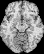
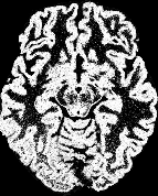
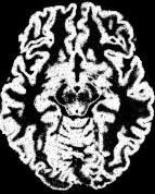
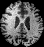
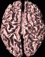
Figure 1 shows an example of applying both standard FCM, AFCM, and FANTASM to a T1-weighted MR image. The soft segmentation results in Figures 1b-c provide models of the partial volume effects present within the image. The FCM result is poor, however, because it does not compensate for the inhomogeneity and noise artifacts within the image. Figures 1e-f show the result the tissue classification result of the brain image into white matter, gray matter, and cerebrospinal fluid (CSF). The AFCM result in Figure 1f compensates for the inhomogeneity artifact within the original image and correctly recovers most of the white matter structure at the bottom of the image, but does not effectively deal with the noise in the image. The FANTASM result of Figure 1g most closely resembles the true classification, shown in Figure 1d. | ||||||||||||||||||||||||||
| ||||||||||||||||||||||||||
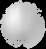
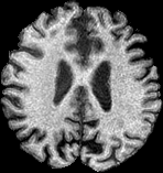
Figure 2 shows how FANTASM can be used to correct for inhomogeneities within an MR image. The inhomogeneities are estimated as a gain field, shown in Figure 2b. The gain field can then be used to correct the shading effect. The right side of Figure 2c is no longer darker than the left side, as it is in Figure 2a. | ||||||||||||||||||||||||||
| ||||||||||||||||||||||||||
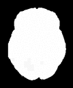
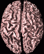
Figure 3 shows the benefits of using FANTASM to reconstruct 3-D surfaces within MR data sets. The surface reconstructed using FANTASM is smoother and more accurate than the surface reconstructed using standard FCM. | ||||||||||||||||||||||||||
| Publications | ||||||||||||||||||||||||||
| ||||||||||||||||||||||||||