Interactive Cortical Editor
Daphne Yu, Chenyang Xu and Jerry L. PrinceOverview
Interactive Cortical Editor (ICE) is a software tool that is aimed to provide a method for the user to edit a previously generated 3D cortical surface model while preserving topology and surface smoothness. With proper modifications and reviews, such models can be highly valuable truth models for evaluting automated cortical surface reconstruction methods. Nevertheless, the highly covoluted nature of the cortical surface poses many difficulties for visualization as well as for many surface editing methods. ICE is set to overcome these major issues.
Introduction
Accurate surface models of the human brain cortex are valuable for brain shape analysis and functional mapping. Despite advances in the methods for cortical surface reconstruction from magnetic resonance images, there has been little progress in the methods for their quantitative validation. Currently, quantitative validation is primarily limited to comparisons with manually picked landmarks from a small subset of the entire surface, and to comparisons with 3-D surface models that were manually traced from the 2-D image slices. These manually traced models are not generally capable of producing the desired topologically correct and smooth surface representation. With new methods developed in ICE, we have manually created a preliminary cortical surface truth model and have used it to obtain error measurements from the entire surface.
Method
To generate a true cortical surface model, we began with an initial surface that was reconstructed from a noise-free and artifact-free simulated brain phantom image acquired from "BrainWeb" [1]. A semi-automatic reconstruction method that employs fuzzy segmentation, isosurface, and active deformation was used and is described in [2]. This reconstructed surface was then carefully reviewed and edited until satisfaction using ICE. This edited surface serves as a true surface model for comparison with other surface models of the brain phantom.
The initial reconstructed surface is a smooth and continuous model of the cortex. These properties should be preserved during surface editing. Additionally, since the cortical surface is highly convoluted, the cortical surface editor must also allow the user to view and edit the deep sulcal surface regions relative to its corresponding location in the 3-D image. Explicitly, the required features in a cortical surface editing tool are the following:
- An interactive visualization environment that allows the user to easily navigate and
cross-reference between the cortical surface and its corresponding image.
- A surface editing method that can move local regions on a 3-D surface while preserving topology and surface smoothness.
|
|
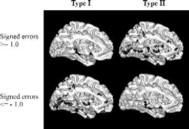
Medial (left image) and right (right image) views of the signed type I and type II errors mapped onto the true (left column) and reconstructed (right column) surfaces respectively. The dark regions indicate where the condition shown is true. Error units are in voxels. |
To address the second problem, ICE processes the user specified surface displacement by our surface-editing algorithm named the Enhanced ChainMail for Surface Meshes (ECSM) algorithm. This algorithm is based on the Enhanced ChainMail algorithm originally developed for fast displacement of volumetric image elements [3]. The ECSM algorithm models edges in a surface mesh as semi-elastic linked elements in a chain. The surface mesh vertices connect these edges, so when a vertex is edited, the displacement stretches or compresses its neighboring edges. In this way, the user specified displacements are propagated efficiently to a 3-D local region in a natural manner that is easily understood by the user. The local region modified using the ECSM algorithm can be further refined by a Gaussian shaping function. This figure shows the results from editing a small region on a spherical surface using various edge elasticity and shaping function parameters.
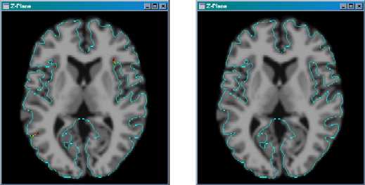
We have found that the combined use of the ECSM algorithm and active deformation was highly effective in correcting most of the erroneous regions that were found in the initial surface. Active deformation is an iterative process that refines the surface via a balance of internal and external forces. It is highly effective for smoothing local edit regions that have been edited using the ECSM algorithm because it regulates the surface elasticity and rigidity through the internal forces. External forces that were computed from the brain image can also be used to assist in guiding the surface to the target location, thus alleviating the amount of editing effort required by the user. This figure shows a cross-section of sulcal regions that is being edited (as indicated by the red and green points) and the result after they have been edited using ICE.
Results
As a preliminary truth model for comparison with a cortical surface that was reconstructed from the same phantom brain model except that noise was added to the image.
For quantitative comparison, we computed two types of errors:
- In Type I, the closest Euclidean distance from each vertex on the
true surface to the reconstructed surface was measured.
- In Type II, the closest Euclidean distance from each vertex on the
reconstructed surface to the true surface was measured.
The following figures illustrate the erroneous regions of the reconstructed surface.
  |
|
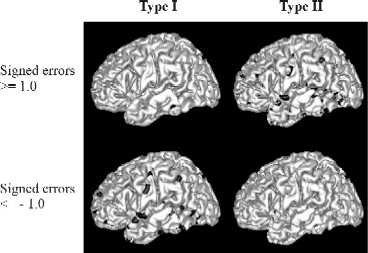
Medial (left image) and right (right image) views of the signed type I and type II errors mapped onto the true (left column) and reconstructed (right column) surfaces respectively. The dark regions indicate where the condition shown is true. Error units are in voxels. |
Conclusion
The ICE software tool is feasible for generating a truth model of the cortical surface
that can be used to obtain error measurements from the entire cortical surface. The ECSM
algorithm used in combination with surface shaping and active deformation surface technique
have also demonstrated to produce the desired changes in a surface. In a preliminary
study, the reconstructed cortical surface have achieved subpixel accuracy in most regions
on the surface even in the presence of image noise. It is important that the truth models
are reviewed by more brain anatomy experts before they are used for further quantitative
validation.
Acknowledgment
We would like to thank Divya Mehra for his efforts in ICE.
This research was supported by NIH/NINDS grant R01NS37747.
Publications
- Yu, D., Xu, C., Prince, J.
"A New Surface Editing Tool for Generating Truth Models of the Human Cortical Surface",
6th International Conference on Function Mapping of the Human Brain, June, 2000.
- Yu, D., Xu, C., Rettmann, M., Pham, D., Prince, J.
"Quantitative Validation of a Deformable Cortical Surface Model",
SPIE International Symposium on Medical Imaging, February 2000.
- Yu, D.
"Quantitative Validation of a Cortical Surface Reconstruction Method",
MSE thesis, The Johns Hopkins University, January 2000.
- Xu, C., Pham, D., Rettmann, M., Yu, D., Prince, J.
"Reconstruction of the Human Cerebral Cortex from Magnetic Resonance Images",
IEEE Transactions in Medical Imaging, 18(6), June 1999.
