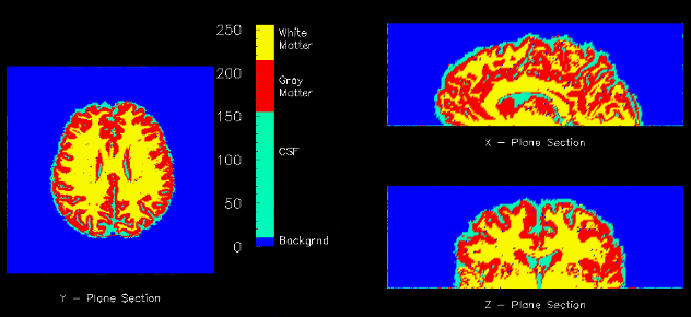
Cross sections of a segmented brain into gray matter, white matter, and cerebrospinal fluid.

Cross sections of a segmented brain into gray matter, white matter, and cerebrospinal fluid.
The goal of image segmentation is to classify the pixels of an image into a certain number of classes that are homogeneous with respect to some characteristic (e.g. intensity, texture). There are many different methods for segmenting an image including clustering, region growing, thresholding, as well as probabilistic methods.
An important area of current research is obtaining more information about brain structure and function. Magnetic Resonance Imaging (MRI )provides a noninvasive, high resolution method of imaging the brain.
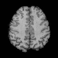
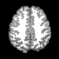
The material of the brain can be categorized into three classes: gray matter, white matter, and cerebrospinal fluid (csf). The ability to segment MR brain images into these three classes is useful in numerous medical applications such as surgical planning, pathological diagnosis, and partial volume averaging correction of PET images. Shown below is a MR image of a brain (axial slice) and its corresponding segmentation. The csf corresponds to the dark gray, gray matter to the medium gray, and white matter to the white.


Two of the more common methods for brain segmentation are the fuzzy c-means clustering algorithm (FCM), and maximum likelihood classification via the expectation maximization (EM) algorithm. Both are iterative optimization algorithms which assign a membership value to each pixel for each class. A hard segmentation can be achieved by assigning each pixel to the class of highest membership.
Current segmentation methods have several drawbacks that require further investigation. First, the problem of magnetic field inhomogeneities is largely ignored by most methods. Inhomogeneites cause pixel intensities to vary spatially across the image. Furthermore, current methods lack a computationally efficient method for modelling context in the image. Because of the large size of the data sets (often 60 slices of 256x256x16bit images), standard Markov random field type algorithms are not computationally viable. Finally, incorporation of topological information into these algorithms could greatly benefit the resulting segmentation.
MC Clark, LO Hall, et al. MRI segmentation using fuzzy clustering techniques. IEEE Engineering in Medicine and Biology, Nov/Dec '94:730-742.
Y Wang and T Lei. A new stochastic model-based image segmentation technique for MR images, IEEE International Conference on Image Processing, ICIP'94, 182-186, 1994.