Finding the Brain Cortex Using Fuzzy Segmentation, Isosurfaces, and Deformable Surface Models
Chenyang Xu, Dzung L. Pham, Maryam E. Rettmann, Daphne Yu, and Jerry L. Prince
Overview
 A method for finding the cortical surface
of the brain from magnetic resonance images using a combination of fuzzy segmentation,
isosurface extraction, and a deformable surface is presented. After MR
images are acquired and preprocessed to remove extracranial tissue, fuzzy membership
functions for gray matter (GM), white matter (WM), and cerebrospinal fluid (CSF)
are computed. An iterative procedure using isosurfaces of filtered WM
membership functions is then used to obtain a topologically correct estimate
of the cortical surface. This estimate forms the initialization of a gradient
vector flow deformable surface, which is then allowed to converge to
the peaks of the GM membership function. The final result is a parameterized
map of the medial layer of the cortex.
A method for finding the cortical surface
of the brain from magnetic resonance images using a combination of fuzzy segmentation,
isosurface extraction, and a deformable surface is presented. After MR
images are acquired and preprocessed to remove extracranial tissue, fuzzy membership
functions for gray matter (GM), white matter (WM), and cerebrospinal fluid (CSF)
are computed. An iterative procedure using isosurfaces of filtered WM
membership functions is then used to obtain a topologically correct estimate
of the cortical surface. This estimate forms the initialization of a gradient
vector flow deformable surface, which is then allowed to converge to
the peaks of the GM membership function. The final result is a parameterized
map of the medial layer of the cortex.
Introduction
Recent advances in medical imaging of the brain allow anatomical information derived from high resolution imaging modalities such as magnetic resonance imaging (MRI) and computed tomography (CT) to be fused with physiological information. These advances have placed a priority on obtaining accurate reconstructions of the cortical surface, not only to provide valuable information on the geometric and anatomical properties of the brain but for other purposes as well. For example, the location of functional activity obtained from positron emission tomography (PET), functional magnetic resonance imaging (fMRI), and other methods can be mapped to the extracted cortical surface, providing a better understanding of brain function and organization. The surfaces can also be warped to other cortical surfaces for the purposes of image registration or atlas labelling. Furthermore, extracted cortical surfaces may be used to study the morphological variability of the brain in aging and among different populations.
Our research is focused on extracting the medial layer surface of the cortex from MR images of the brain. A novel approach is used to provide a proper initialization for a deformable surface based on isosurfaces of a fuzzy segmentation.
Methods and Results
Our technique consists of four major steps: 1) data acquisition and preprocessing, 2) fuzzy segmentation, 3) initial surface estimation, and 4) refinement using a deformable surface. Results of applying each procedure to a sample data set are shown.
- Data acquisition and preprocessing
Data was acquired on a GE Signa 1.5 Tesla MR scanner using a Spoiled GRASS (SPGR) imaging protocol with a pulse repetition time of 35 ms, pulse echo time of 8 ms, and tip angle of 45 degrees. The image size was 256x240, zero padded to 256x256 with pixel dimensions of 0.9375x0.9375 mm. 124 axial slices were acquired with a slice thickness of 1.5mm.
The acquired images were stacked to form a volume and then the extracranial tissue removal was accomplished using a semi-automated software package developed by Christos Davatzikos and Jerry Prince.
- Fuzzy segmentation
The preprocessed brain volume is segmented by applying fuzzy c-means (FCM) algorithm, resulting in fuzzy membership functions for GM, WM, and CSF tissue classes. The resulting membership functions for one slice in the brain volume are shown in Fig. 1.
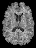


(a) (b) (c) (d) Fig. 1. (a) a sample slice from acquired MRI data set. Membership functions: (b) gray matter (GM), (c) white matter (WM), and (d) cerebrospinal fluid (CSF). - Initial estimation of the
cortical surface
 In order to use a deformable
surface to extract the surface of the cortex, a proper initialization is
required. The initialization must be sufficiently close and isomorphic (i.e.,
have an equivalent topology) to the medial layer surface of the GM.
In order to use a deformable
surface to extract the surface of the cortex, a proper initialization is
required. The initialization must be sufficiently close and isomorphic (i.e.,
have an equivalent topology) to the medial layer surface of the GM.
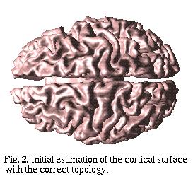
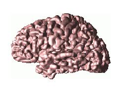
In our approach, we use isosurface to provide initial estimation of the cortical surface. However, isosurface in general does not has constraint on topology and sensitive to noise. An automatic iterative procedure which involves median filtering and isosurface is applied to generate topologically correct cortical surface estimation. The result of this procedure is shown in Fig. 2.
- Refinement of the cortical
surface
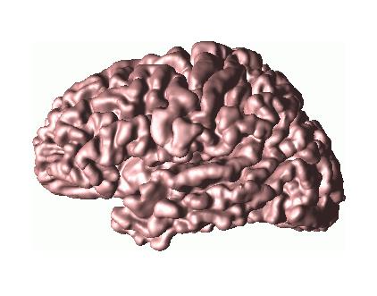
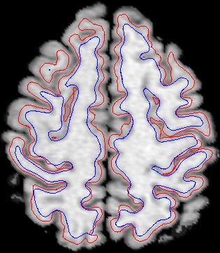
After obtaining an initial estimate of the cortical surface which is topologically correct, the surface requires refinement. The initial surface is a smoothed version of the GM-WM interface. Using deformable surfaces, it is possible to have this initial surface move towards the desired surface (i.e., the medial layer surface of the GM). The main problem here lies in defining the external forces. Recently, we have proposed a new type of external force for active contour and deformable surfaces called Gradient Vector Flow (GVF). GVF has many advantages over the traditional external forces. Detailed comparisons between GVF and traditional external forces could be found in [4,5]. The property of GVF makes it a suitable choice for our cortical surface refinement. Fig. 3 shows the result of the final deformed surface on our sample data set using GVF based deformable surface. A cross section of the initial and final deformable surface overlaid on top of the MRI image is shown in Fig. 4.
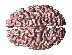
(a) (b) Fig. 3. Final GVF deformable surface. (a) Top view and (b) left view. 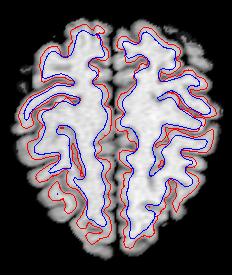
Fig. 4. Cross section of the initial and final deformable surface overlaid on top of the MRI image. Initial surface is plotted as blue and final surface is plotted as red.

Differential geometry and partial flattening
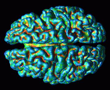
Once the cortex has been extracted, differential geometric quantities may be computed. Differential geometry provides a natural mathematical framework to study the cortical surface geometry. Among many geometric quantities defined for the surface, curvature information is the most valuable since it quantifies the structure of the sulci and gyri, thus providing the basis for scientific and comparative studies.
Fig. 5 shows two views of the mean curvature, plotted on the extracted cortical surface of our sample data set. Red in the mean curvature figure stands for positive mean curvature values, green stands for zero, and blue stands for negative mean curvature values. From the figures, we see that sulci are described by red central ``skeletons'' surrounded by blue regions. These skeletons, which are in the interior of the brain, correspond to the ``roots'' of the sulci, while the blue regions are the ``lips'' of the sulci on the surface of the brain.
 |
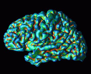 |
| (a) | (b) |
| Fig. 5. Mean curvature plotted onto the final GVF deformable surface. (a) Top view and (b) left view. | |
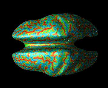
Once the cortical surface is extracted, a common practice in brain mapping community is to let the cortical surface undergo partial flattening. Partial flattening has the advantage of maintaining the overall shape of the original cortical surface while revealing embedded sulci. Partial flattening can be achieved by simply subjecting the deformable surface model to only internal forces. Fig. 6 shows the mean curvature of the original cortical surface plotted on partial flattened surface. The locations and geometric details of the sulci, as revealed by the mean curvature calculations, are even more strongly apparent in this figure, since we can see the entire surface. It is this type of information that many believe will allow further advancement in brain mapping using medical imaging.
 |
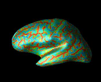 |
| (a) | (b) |
| Fig. 6. Mean curvature plotted onto the partial flattened cortical surface. (a) Top view and (b) left view. | |
Conclusion
The presented method for finding
the cortical surface in MR data has the potential to preserve even the deepest
folds in the cortex using deformable surface models. Several possibilities exist,
however, for further improvement. For example, more sophisticated segmentation
algorithms are available which take into account the spatial dependence of image
intensities and MRI artifacts, such as intrascan intensity inhomogeneities.
In order to take full advantage of our method, however, a fuzzy segmentation
output is required. Also, an improvement in the initial cortical surface may
be obtained by using a different isosurface threshold, or by computing an isosurface
on a function of the membership functions.
Publications
- C. Xu and J. L. Prince, "A Generalized Gradient Vector Flow for Active Contour Models," Proc. Conf. Inf. Sci. Sys., The Johns Hopkins Univ., pp. 885-890, March 19-21, 1997
- C. Xu, D.L. Pham, and J.L. Prince, "Finding the Brain Cortex Using Fuzzy Segmentation, Isosurfaces, and Deformable Surface Models", Proceedings of the XVth International Conference on Information Processing in Medical Imaging (IPMI'97), 399-404, June 1997.
- C. Xu, D. L. Pham, M. E. Etemad, D. N. Yu, and J. L. Prince, "Reconstruction of the Central Layer of the Human Cerebral Cortex from MR images," in Proc. of the First International Conference on Medical Image Computing and Computer Assisted Interventions (MICCAI'98), pp. 482-488, 1998.
- C. Xu, D. L. Pham, and J. L. Prince, "Reconstruction of the Human Cortical Surface from MR Images", 4th International conference on Functional Mapping of the Human Brain, June 7-12, 1998, Montreal, Quebec, Canada; NeuroImage, vol. 7, no. 4, pg. 715, May 1998.
-
C. Xu, D. L. Pham, M. E. Rettmann, D. N. Yu, and J. L. Prince, "Reconstruction of the Human Cerebral Cortex from Magnetic Resonance Images," IEEE Transactions on Medical Imaging, 18(6), pp. 467-480, June, 1999.
-
C. Xu and J.L. Prince, "Gradient Vector Flow: A New External Force for Snakes", IEEE Proc. Conf. on Comp. Vis. Patt. Recog. (CVPR'97). 66-71, June 1997.
-
C. Xu and J.L. Prince, "Snakes, Shapes, and Gradient Vector Flow", IEEE Transactions on Image Processing, 359-369, March, 1998 (JHU-ECE TR96-15). Click here to see a GVF demo.
- C. Xu, M. E. Etemad, D. Yu, D. L. Pham, and J. L. Prince, "A Spherical Map for Cortical Geometry", 4th International Conference on Function Mapping of the Human Brain, June 7-12, 1998, Montreal, Quebec, Canada; NeuroImage, vol. 4, no. 4, pg. 734, May 1998.
- C. Xu and J. L. Prince, "Generalized Gradient Vector Flow External Forces for Active Contours", Signal Processing, vol. 71, no. 2, pp. 131-139, December 1998.
-
D. L. Pham and J. L. Prince, "An Adaptive Fuzzy C-Means Algorithm for Image Segmentation in the Presence of Intensity Inhomogeneities" Pattern Recognition Letters, vol. 20, no. 1, pp. 57-68, 15-January, 1999.
- C. Xu, D. L. Pham, M. E. Rettmann, D. N. Yu, and J. L. Prince, "Reconstruction of the human cerebral cortex from magnetic resonance images", IEEE Trans. Medical Imaging, vol. 18, no. 6, pp. 467-480, June 1999.
- C. Xu, D. L. Pham, and J. L. Prince "Image Segmentation Using Deformable Models", in Handbook of Medical Imaging: Volume 2. Medical Image Processing and Analysis, eds. M. Sonka and J. M. Fitzpatrick, SPIE Press, pp.129-174, 2000.
- C. Xu and J. L. Prince , "Gradient Vector Flow Deformable Models", in Handbook of Medical Image Processing and Analysis, ed. Isaac N. Bankman, Academic Press, pp.159-170, 2000.
- X. Han, M. E. Rettmann, C. Xu, and J. L. Prince, "Automatic segmentation editing for cortical surface reconstruction," Proc. SPIE Medical Imaging, Conf. 4322, Paper 22, San Diego, February 2001.
- D.
Tosun and J. L. Prince,
"Hemispherical map for the human brain cortex," Proc. SPIE Medical
Imaging, Conf. 4322, Paper 31, San Diego, February 2001.
