Cardiac Material Markers from Tagged MR Images
Williams S. Kerwin and Jerry L. Prince
Overview
We have developed a technique for tracking the motion of the left ventricle of the heart during contraction. The technique uses a sequence of magnetic resonance images and an established imaging method known as "tagging". We use tagged MR data in a unique method that produces 3-D motion estimates over a discrete set of points (so-called material points) in the wall of the left ventricle. The method relies on a multi-step process that includes use of thin-plate splines to interpolate data between image planes and an iterative, alternating projections algorithm for locating the tracked material points. The details of the method are presented below.
 An
example of the resulting data is presented at the left (click on the image to
view an mpeg movie). The movie shows the position of some 254 material
points at ten time frames during the contraction of the left ventricle.
The points are viewed down the center of the left ventricle. Watch for
the characteristic twisting of the left ventricle. Such data can be used
for visualization of heart function or for developing models of heart motion.
We are also working to extract clinically useful information for the early detection
and assessment of ischemic heart disease.
An
example of the resulting data is presented at the left (click on the image to
view an mpeg movie). The movie shows the position of some 254 material
points at ten time frames during the contraction of the left ventricle.
The points are viewed down the center of the left ventricle. Watch for
the characteristic twisting of the left ventricle. Such data can be used
for visualization of heart function or for developing models of heart motion.
We are also working to extract clinically useful information for the early detection
and assessment of ischemic heart disease.
Introduction
Measurement of heart wall motion
during contraction is a fundamental challenge in the study of cardiac mechanics.
Motion measurement has the potential to be used as a diagnostic tool for assessment
of heart disease and as a theoretical tool for physiological analysis.
Past approaches for measuring heart motion can be divided into two types. One
type tracks the movement of a set of points - called material markers - which
are either implanted or naturally occuring landmarks within the heart.
The second type of approach uses tagged magnetic resonance imaging techniques
to estimate the displacement field for all points within the heart. Here
we present a technique that uses the standard MR tagging protocol, but measures
motion at a discrete set of material points as in the material marker methods.
We refer to the resulting motion data as "MR markers". This technique
may be used to compare the merits of MR tagging to material marker techniques,
or to provide data to existing analysis routines intended for use with material
marker data.
Method
 We
use standard tagged MRimages, where "tagging" is a method that aids in tracking
motion of objects using a series of magnetic resonance images. Tags appear
as dark regions in the images that move with an object as it moves. Tagging
is particularly valuable in cardiac imaging because the tissue in the walls
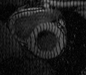
of the heart provides few natural features for motion tracking. An example
of a tagged heart image is shown at left, where the ring shaped object is the
wall of the left ventricle. The tag pattern used was originally a set
of parallel stripes and this image was taken after heart motion had deformed
the stripes into the bent lines you see here. Click on the image to see
a 10 frame mpeg movie demonstrating the deformation of the tag lines from their
original straight configuration.
We
use standard tagged MRimages, where "tagging" is a method that aids in tracking
motion of objects using a series of magnetic resonance images. Tags appear
as dark regions in the images that move with an object as it moves. Tagging
is particularly valuable in cardiac imaging because the tissue in the walls
of the heart provides few natural features for motion tracking. An example
of a tagged heart image is shown at left, where the ring shaped object is the
wall of the left ventricle. The tag pattern used was originally a set
of parallel stripes and this image was taken after heart motion had deformed
the stripes into the bent lines you see here. Click on the image to see
a 10 frame mpeg movie demonstrating the deformation of the tag lines from their
original straight configuration.
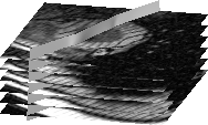
Although "tag lines" is the normal term for these induced features, tags generally consist of sheets of dark (when imaged) tissue. Tag lines are the intersection of these "tag surfaces" with image planes. This concept is illustrated in the next image below, which shows a single tag surface intersecting a set of images. The tag surface contributes one tag line to each of the image planes. A standard tagged MRI data set consists of many such tag surfaces arranged in a regular 3-D grid. The contracting heart deforms this regular grid and from such deformation, motion information is extracted. To illustrate this point, click on the second image below to view a movie showing the deforming grid.
 |
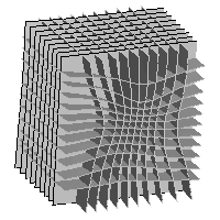 |
Our complete method is a three step process consisting of grid reconstruction, surface intersection identification, and segmentation. Grid reconstruction must be performed because the position of the tag surfaces is only known within image planes. Between image planes, the tag surfaces are estimated using thin-plate splines, which individually interpolate each surface. The complete reconstructed grid (as depicted above) is the combination of the thin-plate spline surfaces. Next, all grid intersection points are identified using an iterative, alternating-projections algorithm. In the publications listed, we offer a proof that this algorithm converges to the unique intersection point of any three surfaces. The identified points are then the nodes of a regular 3-D grid as depicted in the next image. Finally, the points are segmented to identify only those within the wall of the left ventricle. The result is a cup shaped set of MR markers as shown in the second image below.
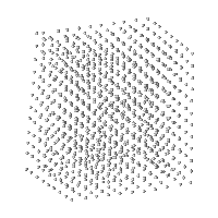 |
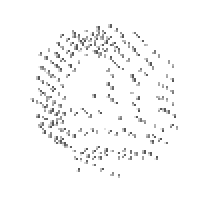 |
Results
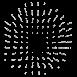
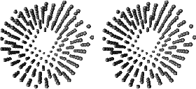
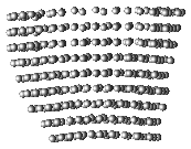
This method has been applied to several data sets. Here we present a typical example. The tagged MRI protocol was applied to a healthy human volunteer. The initial distance between tag surfaces was 6mm and images were taken every 32.5msec for ten times during contraction of the heart. After applying our algorithm, 254 MR markers were identified within the wall of the left ventricle. Images of the results are depicted below. The first image is a stereo pair viewing the left ventricle down the long axis (from base toward apex). This image can be viewed in 3-D by focusing your left eye on the left image and right eye on the right image. A 3-D movie is also available by clicking on the image. Viewing this image in 3-D is useful for demonstrating the distribution of the markers in space. Finally, the last image is a side view of the MR markers with the free wall of the left ventricle on the left. Click on the image to view a movie version of this image, which will demonstrate the vertical shortening of the left ventricle during contraction.
Publications
- W.S. Kerwin and J.L. Prince, ``Generating 3-D Cardiac Material Markers Using Tagged MRI,'' Proc. Information Processing in Medical Imaging (IPMI), Berlin: Springer-Verlag, pp. 313-326, June 1997. [request hard copy]
- W. S. Kerwin and J. L. Prince, "Recursive Filtering and Interpolation Algorithm for Function Sequences of Any Dimension", Proc. Image and Multidim. Dig. Sig. Proc., pp.239-242, Alpbach, July 1998.
- W. S. Kerwin and J. L. Prince, "MR Tag Surface Tracking Using a Spatio-temporal Filter/Interpolator", Proceedings of IEEE Int. Conf. Image Proc., vol. 1, pp. 699-703, Chicago, October 1998.
- W. S. Kerwin and J. L. Prince, "Cardiac Material Markers from Tagged MR Images," Medical Image Analysis, vol. 2, issue 4, pp. 339-353, 1998.
- W. S. Kerwin and J. L. Prince, "Tracking MR tag surfaces using a spatio-temporal filter and interpolator," Int'l Journal of Imaging Systems and Technology, vol. 10, pp. 128-142, 1999.
- W. S. Kerwin and J. L. Prince, "The Kriging Update Model and Recursive Space-Time Function Estimation," IEEE Trans. on Signal Processing, vol. 47, no. 11, pp. 2942-2952, November 1999.
- W. S. Kerwin and J. L. Prince,
"Kriging Filters for Space Time Interpolation," Advances in Imaging
and Electron Physics, invited by Dr. Peter Hawkes, 124:139-193, November 2002.
