Difference between revisions of "JohnVisualizations"
Jump to navigation
Jump to search
(another image) |
|||
| (3 intermediate revisions by one other user not shown) | |||
| Line 1: | Line 1: | ||
| − | <meta name="title" content="Data Visualization"/> | + | <!-- <meta name="title" content="Data Visualization"/> --> |
{{h2|Data Visualization}} | {{h2|Data Visualization}} | ||
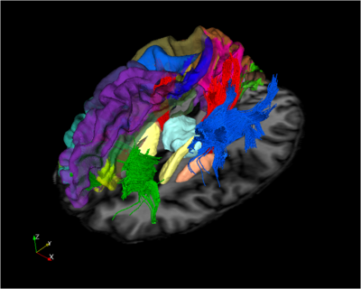
| − | [[Image:ObliqueCortSubcortTracts.png|left|thumb|400px|A 3D rendering of a labeled brain | + | {|class="wikitable" style="width: 70%; text-align: top;" |
| + | |- | ||
| + | | [[Image:ObliqueCortSubcortTracts.png|left|thumb|400px|A 3D rendering of a labeled brain | ||
surface, overlaid on an MR image. Sub-cortical structures and three white-matter tracts are visible.]] | surface, overlaid on an MR image. Sub-cortical structures and three white-matter tracts are visible.]] | ||
| + | || | ||
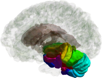
| + | | [[Image:CerSlices_rnder_20120810.png|left|thumb|400px|A rendering of the cerebellar lobules with the cerebral cortex and subcortical structures.]] | ||
| + | |- | ||
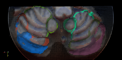
| + | | [[Image:Cer_FieldAndMpr_wMultisurface.png|left|thumb|400px|A vector field and a cut-away of a labeled cerebellum surface overlaid on an MRI slice of the human cerebellum.]] | ||
| + | || | ||
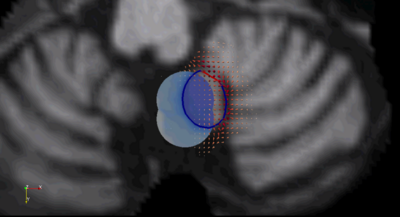
| + | | [[Image:Movie_lobule9_thumbnail.png|left|thumb|400px|[http://iacl.ece.jhu.edu/~john/mgdm_lobule9_field_intensitySpeed.avi A movie of the evolution of cerebellar lobule 9 according to boundary-specific speeds. The vector field shown by red glyphs influences the red portion of the boundary, while the blue spotlights influence the blue portion of the object boundary.] ]] | ||
| + | |- | ||
| + | |} | ||
| + | <br> | ||
| − | [[Image: | + | {{h4|Cerebellum Shape}} |
| + | {|class="wikitable" style="width: 520px; text-align: center;" | ||
| + | |- | ||
| + | |[[Image:Anura meanSdfDiff axl 20100930.png | 250px]] || [[Image:Anura meanSdfDiff cor 20100930.png | 250px]] | ||
| + | |- | ||
| + | | [[Image:Anura meanSdfDiff sag 20100930.png | 250px]] || [[Image:Anura meanSdfDiff refRndr 20100930.png | 250px]] | ||
| + | |- | ||
| + | |} | ||
