Difference between revisions of "MSChallenge"
(Cleaned up the current leader board.) |
(Completing table.) |
||
| Line 42: | Line 42: | ||
|- | |- | ||
| 3 | | 3 | ||
| − | | ''MV-CNN''<br /> | + | | ''MV-CNN''<br />A. Birenbaum & H. Greenspan<br />'''Multi-View Convolutional Neural Networks'''<br />{{pub|doi=10.1007/978-3-319-46976-8_7|period=}} |
| | | | ||
| 90.070 | | 90.070 | ||
| Line 70: | Line 70: | ||
|- | |- | ||
| 7 | | 7 | ||
| − | | ''Lesion-TOADS''<br />N. Shiee, P.-L. Bazin, & D.L. Pham<br /> | + | | ''Lesion-TOADS''<br />N. Shiee, P.-L. Bazin, A. Ozturk, D. S. Reich, P. A. Calabresi, & D. L. Pham<br />'''A topology-preserving approach to the segmentation of brain images with multiple sclerosis lesions'''<br />{{pub|doi=10.1016/j.neuroimage.2009.09.005|pubmed=19766196|pmcid=2806481|period=}} |
| | | | ||
| 88.465 | | 88.465 | ||
| Line 84: | Line 84: | ||
|- | |- | ||
| 9 | | 9 | ||
| − | | ''MORF''<br /> | + | | ''MORF''<br />A. Jog, A. Carass, D.L. Pham, & J.L. Prince<br />'''Multi-Output Random Forests for Lesion Segmentation in Multiple Sclerosis'''<br />{{pub|doi=10.1117/12.2082157|pubmed=27695155|pmcid=5041594|period=}} |
| | | | ||
| 87.917 | | 87.917 | ||
Revision as of 21:51, 18 November 2016
<meta name="title" content="MS Challenge" />
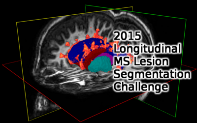
The 2015 Longitudinal Multiple Sclerosis Lesion Segmentation Challenge
I. Introduction
The Longitudinal MS Lesion Segmentation Challenge will be conducted at the 2015 International Symposium on Biomedical Imaging in New York, NY, April 16-19. Competing teams will apply their automatic lesion segmentation algorithms to MR neuroimaging data acquired at multiple time points from MS patients. Algorithms will be evaluated against manual segmentations from multiple raters in terms of their segmentation accuracy and ability to track lesion evolution.
Registration for the Challenge is now closed. 34 Teams initially registered for the Challenge coming from 15 different countries, representing 27 different institutions/universities. Congratulations to Team IIT Madras (First Prize), Team PVG_1 (Second Prize), and Team IMI (Third Prize and Efficiency Prize)!
Current Leaderboard
| Leaderboard | |||
| Ranking | Method Name Authors Paper Title Paper Link(s) |
Website Score | |
| 1 | Team PVG One A. Jesson & T. Arbel Hierarchical MRF and Random Forest Segmentation of MS Lesions and Healthy Tissues in Brain MRI (PDF) |
90.698 | |
| 2 | Team IMI O. Maier & H. Handels MS-Lesion Segmentation in MRI with Random Forests (PDF) |
90.283 | |
| 3 | MV-CNN A. Birenbaum & H. Greenspan Multi-View Convolutional Neural Networks |
90.070 | |
| 4 | Team VISAGES GCEM L. Catanese, O. Commowick, & C. Barillot Automatic Graph Cut Segmentation of Multiple Sclerosis Lesions (PDF) |
89.807 | |
| 5 | Team IIT Madras S. Vaidya, A. Chunduru, R. Muthuganapathy, & G. Krishnamurthi Longitudinal Multiple Sclerosis Lesion Segmentation using 3D Convolutional Neural Networks (PDF) |
89.159 | |
| 6 | Team MS*metrix* S. Jain, D.M. Sima, & D. Smeets Automatic Longitudinal Multiple Sclerosis Lesion Segmentation (PDF) |
88.744 | |
| 7 | Lesion-TOADS N. Shiee, P.-L. Bazin, A. Ozturk, D. S. Reich, P. A. Calabresi, & D. L. Pham A topology-preserving approach to the segmentation of brain images with multiple sclerosis lesions |
88.465 | |
| 8 | Team CMIC F. Prados, M.J. Cardoso, N. Cawley, O. Ciccarelli, C.A.M. Wheeler-Kingshott, & S. Ourselin Multi-Contrast PatchMatch Algorithm for Multiple Sclerosis Lesion Detection (PDF) |
88.009 | |
| 9 | MORF A. Jog, A. Carass, D.L. Pham, & J.L. Prince Multi-Output Random Forests for Lesion Segmentation in Multiple Sclerosis |
87.917 | |
| 10 | Team TIG-BF C.H. Sudre, M.J. Cardoso, & S. Ourselin Model Selection Propagation for Application on Longitudinal MS Lesion Segmentation (PDF) |
87.376 | |
| 11 | Team CRL X. Tomas-Fernandez & S.K. Warfield Model of Population and Subject (MOPS) Segmentation (PDF) |
87.017 | |
| 12 | Team DIAG M. Ghafoorian & B. Platel Convolution Neural Networks for MS Lesion Segmentation (PDF) |
86.916 | |
| 13 | Team TIG C.H. Sudre, M.J. Cardoso, & S. Ourselin Model Selection Propagation for Application on Longitudinal MS Lesion Segmentation (PDF) |
86.436 | |
| 14 | Team VISAGES DL H. Deshpande, P. Maurel, & C. Barillot Sparse Representations and Dictionary Learning Based Longitudinal Segmentation of Multiple Sclerosis Lesions (PDF) |
86.068 | |
| 15 | Team BAUMIP L.O. Iheme & D. Unay Automatic White Matter Hyperintensity Segmentation using FLAIR MRI (PDF) |
84.140 | |
II. Data
The overall data will be composed of three parts: 1) Training data consisting of longitudinal images from 5 patients; 2) Test data 1 consisting of longitudinal images from 10 patients; and 3) Test data 2 consisting of longitudinal images from 5 patients. Only the training data will include manual delineations when it is released to teams. These delineations will be performed by at least 2 trained raters. Test data 1 will be released in advance of the challenge day and test data 2 will be released on or shortly before the challenge day to discourage patient-specific tuning of the algorithms and computationally inefficient approaches.
Each longitudinal dataset will include T1-weighted, T2-weighted, PD-weighted, and T2-weighted FLAIR MRI with 3-5 time points acquired on a 3T MR scanner. T1-weighted images will have approximately a 1mm cubic voxel resolution, while the other scans will be 1mm in plane with 3mm sections. Accounting for the multiple time points, this constitutes approximately 80 individual data sets. To minimize the dependency of the results on registration performance and brain extraction, all images will be provided already rigidly co-registered to the baseline T1-weighted image with automatically computed skull stripping masks that may optionally be used by the teams.
The training and test data will continue to be made publicly available following the challenge, similar to the MICCAI 2008 Lesion Challenge data. Manual delineations on the test data will not be made available to the public but the organizers will provide evaluation results for any submitted segmentations. A public website for disseminating the data is currently in development.
III. Evaluation
Here are some initial guidelines.
The evaluation software can be downloaded as Matlab Dot M files (339KB) and as a compiled Matlab executable (46MB). The evaluation metric currently include: Dice Overlap, Jaccard Overlap, PPV (positive predictive value), TPR (sensitivity, voxel based), LTPR (lesion TPR based on lesion count), LFPR (lesion FPR based on lesion count), Volume Difference, Surface Difference, Segmentation Volume, Volume Change Correlation, New lesion detection TPR, and New lesion detection FPR.
VII. Organizers
Primary Organizer:
Dzung Pham, Center for Neuroscience and Regenerative Medicine, Henry M. Jackson Foundation for the Advancement of Military Medicine, Bethesda, MD
Organizing committee members:
Pierre-Louis Bazin, Department of Neurophysics, Max Planck Institute, Leipzig, Germany
Aaron Carass, Department of Electrical and Computer Engineering, Johns Hopkins University, Baltimore, MD
Peter Calabresi, Department of Neurology, Johns Hopkins University, Baltimore, MD
Ciprian Crainiceanu, Department of Biostatistics, Johns Hopkins University, Baltimore, MD
Lotta Ellingsen, Department of Electrical and Computer Engineering, University of Iceland, Reykjavik, Iceland
Qing He, Center for Neuroscience and Regenerative Medicine, Henry Jackson Foundation, Bethesda, MD
Jerry Prince, Department of Electrical and Computer Engineering, Johns Hopkins University, Baltimore, MD
Daniel Reich, Translational Neuroradiology Unit, National Institute of Neurological Disorders and Stroke, National Institutes of Health, Bethesda, MD
Snehashis Roy, Center for Neuroscience and Regenerative Medicine, Henry Jackson Foundation, Bethesda, MD
VIII. Funding support
This challenge is supported in part by a grant from the National Institute of Neurological Disorders and Stroke (NINDS R01 NS070906). Funding for prizes is supported by the National Multiple Sclerosis Society.


