Difference between revisions of "Main Page"
Jump to navigation
Jump to search
(Correction.) |
(Trying without the spacing.) |
||
| Line 5: | Line 5: | ||
<ul class="bss"> | <ul class="bss"> | ||
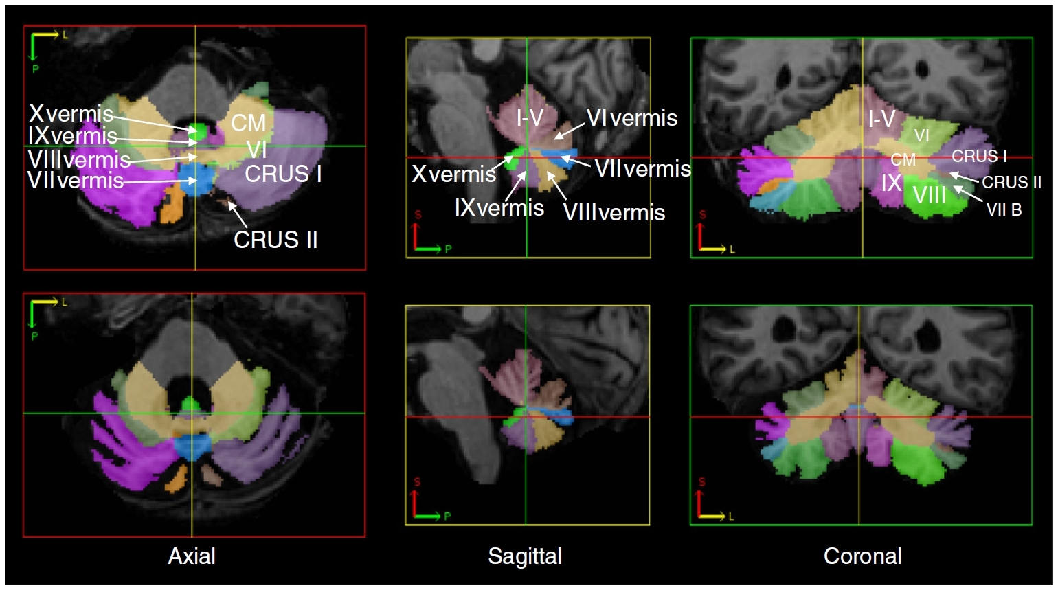
<li>[[File:2016-NI-Yang.jpg|link=http://dx.doi.org/10.1016/j.neuroimage.2015.09.032|center]]<p class="bss-caption text-center">Z. Yang, C. Ye, J.A. Bogovic, A. Carass, B.M. Jedynak, S.H. Ying, and J.L. Prince,<span> </span><b>"Automated Cerebellar Lobule Segmentation with Application to Cerebellar Structural Analysis in Cerebellar Disease"</b>,<span> </span><i>NeuroImage</i>,<span> </span>127:435-444,<span> </span>2016.<span> </span>[http://dx.doi.org/10.1016/j.neuroimage.2015.09.032 (doi)]</p></li> | <li>[[File:2016-NI-Yang.jpg|link=http://dx.doi.org/10.1016/j.neuroimage.2015.09.032|center]]<p class="bss-caption text-center">Z. Yang, C. Ye, J.A. Bogovic, A. Carass, B.M. Jedynak, S.H. Ying, and J.L. Prince,<span> </span><b>"Automated Cerebellar Lobule Segmentation with Application to Cerebellar Structural Analysis in Cerebellar Disease"</b>,<span> </span><i>NeuroImage</i>,<span> </span>127:435-444,<span> </span>2016.<span> </span>[http://dx.doi.org/10.1016/j.neuroimage.2015.09.032 (doi)]</p></li> | ||
| − | |||
| − | |||
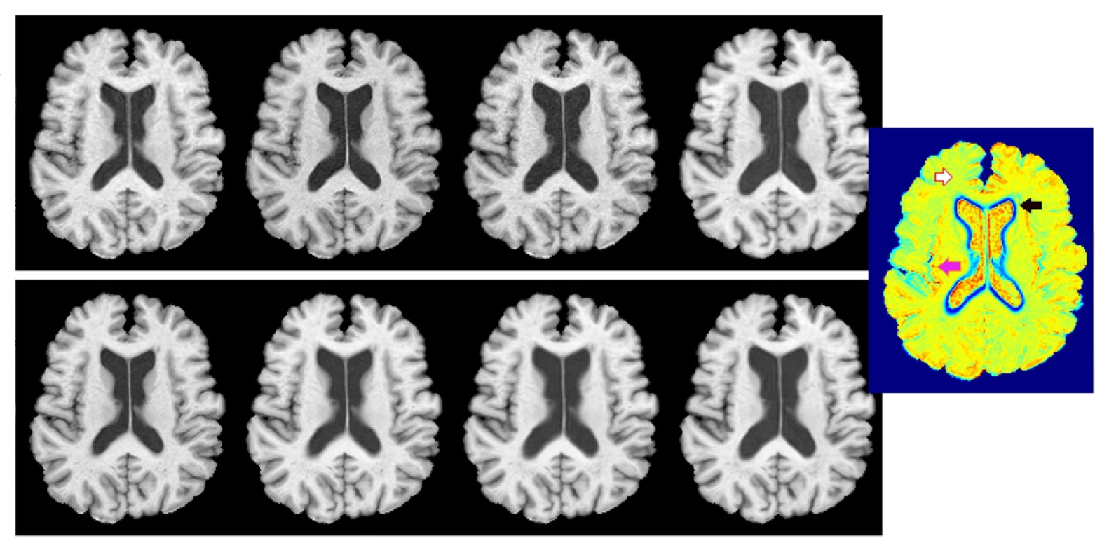
<li>[[File:2016-NIC-Roy.png|link=http://dx.doi.org/doi:10.1016/j.nicl.2016.02.005|center]]<p class="bss-caption text-center">S. Roy, A. Carass, J. Pacheco, M. Bilgel, S.M. Resnick, J.L. Prince, and D.L. Pham,<span> </span><b>"Temporal filtering of longitudinal brain magnetic resonance images for consistent segmentation"</b>,<span> </span><i>NeuroImage: Clinical</i>,<span> </span>11:264-275, 2016,<span> </span>[http://dx.doi.org/doi:10.1016/j.nicl.2016.02.005 (doi)]</p></li> | <li>[[File:2016-NIC-Roy.png|link=http://dx.doi.org/doi:10.1016/j.nicl.2016.02.005|center]]<p class="bss-caption text-center">S. Roy, A. Carass, J. Pacheco, M. Bilgel, S.M. Resnick, J.L. Prince, and D.L. Pham,<span> </span><b>"Temporal filtering of longitudinal brain magnetic resonance images for consistent segmentation"</b>,<span> </span><i>NeuroImage: Clinical</i>,<span> </span>11:264-275, 2016,<span> </span>[http://dx.doi.org/doi:10.1016/j.nicl.2016.02.005 (doi)]</p></li> | ||
| − | |||
| − | |||
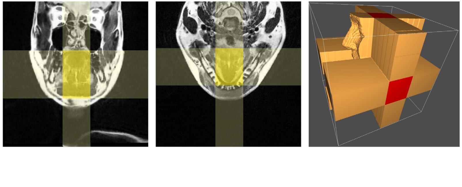
<li>[[File:2015-MIA-Ibragimov.jpg|link=http://dx.doi.org/10.1016/j.media.2014.11.006|center]]<p class="bss-caption text-center">B. Ibragimov, J.L. Prince, E.Z. Murano, J. Woo, M. Stone, B. Likar, F. Pernuš, and T. Vrtovec,<span> </span><b>"Segmentation of tongue muscles from super-resolution magnetic resonance images"</b>,<span> </span><i>Medical Image Analysis</i>,<span> </span>20(1):198-207,<span> </span>2015.<span> </span>[http://dx.doi.org/10.1016/j.media.2014.11.006 (doi)]</p></li> | <li>[[File:2015-MIA-Ibragimov.jpg|link=http://dx.doi.org/10.1016/j.media.2014.11.006|center]]<p class="bss-caption text-center">B. Ibragimov, J.L. Prince, E.Z. Murano, J. Woo, M. Stone, B. Likar, F. Pernuš, and T. Vrtovec,<span> </span><b>"Segmentation of tongue muscles from super-resolution magnetic resonance images"</b>,<span> </span><i>Medical Image Analysis</i>,<span> </span>20(1):198-207,<span> </span>2015.<span> </span>[http://dx.doi.org/10.1016/j.media.2014.11.006 (doi)]</p></li> | ||
| − | |||
| − | |||
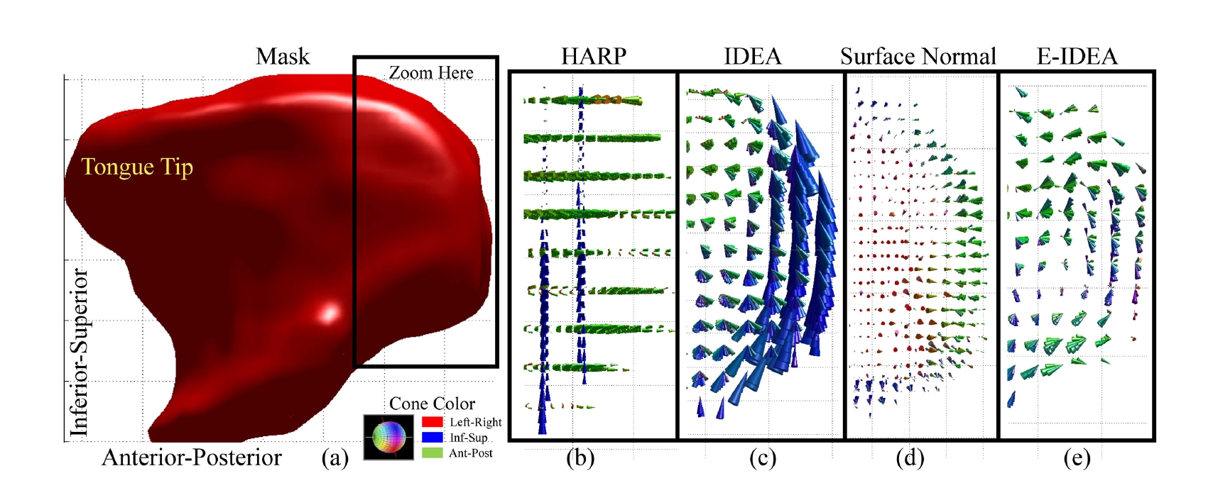
<li>[[File:2013-MICCAI-Xing.jpg|link=http://dx.doi.org/10.1007/978-3-642-40760-4_6|center]]<p class="bss-caption text-center">F. Xing, J. Woo, E.Z. Murano, J. Lee, M. Stone, and J.L. Prince,<span> </span><b>"3D Tongue Motion from Tagged and Cine MR Images"</b>,<span> </span><i>16th International Conference on Medical Image Computing and Computer Assisted Intervention</i> (MICCAI 2013), Nagoya, Japan, September 22-26, 2013.<span> </span>[http://dx.doi.org/10.1007/978-3-642-40760-4_6 (doi)]</p></li> | <li>[[File:2013-MICCAI-Xing.jpg|link=http://dx.doi.org/10.1007/978-3-642-40760-4_6|center]]<p class="bss-caption text-center">F. Xing, J. Woo, E.Z. Murano, J. Lee, M. Stone, and J.L. Prince,<span> </span><b>"3D Tongue Motion from Tagged and Cine MR Images"</b>,<span> </span><i>16th International Conference on Medical Image Computing and Computer Assisted Intervention</i> (MICCAI 2013), Nagoya, Japan, September 22-26, 2013.<span> </span>[http://dx.doi.org/10.1007/978-3-642-40760-4_6 (doi)]</p></li> | ||
| − | |||
| − | |||
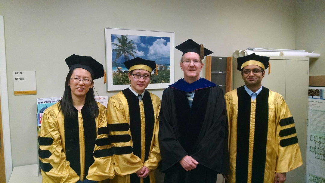
<li>[[File:IMG_20160517_120218495_25.jpg|link=News|center]]<p class="bss-caption text-center">Drs. Yang, Xing, and Jog celebrate receiving their PhDs with Jerry L. Prince.</p></li> | <li>[[File:IMG_20160517_120218495_25.jpg|link=News|center]]<p class="bss-caption text-center">Drs. Yang, Xing, and Jog celebrate receiving their PhDs with Jerry L. Prince.</p></li> | ||
| − | |||
| − | |||
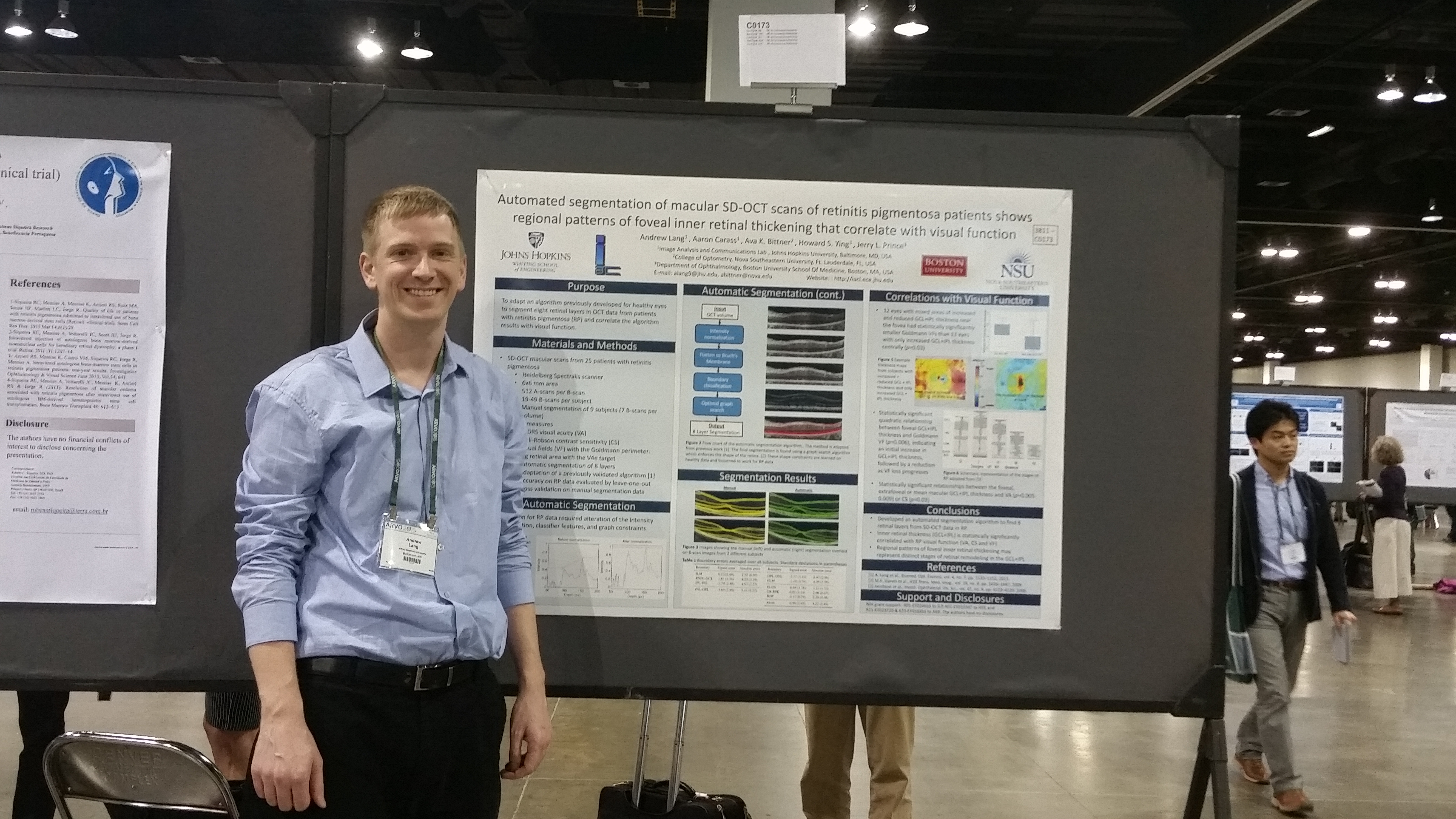
<li>[[File:Andrew_Lang_ARVO2015.jpg|link=News|center]]<p class="bss-caption text-center">Dr. Andrew Lang presented his poster "Automated segmentation of macular SD-OCT scans of retinitis pigmentosa patients shows regional patterns of foveal inner retinal thickening that correlate with visual function" at ARVO 2015 in Denver, Colorado.</p></li> | <li>[[File:Andrew_Lang_ARVO2015.jpg|link=News|center]]<p class="bss-caption text-center">Dr. Andrew Lang presented his poster "Automated segmentation of macular SD-OCT scans of retinitis pigmentosa patients shows regional patterns of foveal inner retinal thickening that correlate with visual function" at ARVO 2015 in Denver, Colorado.</p></li> | ||
| − | |||
| − | |||
</ul> | </ul> | ||
</div> | </div> | ||
</center> | </center> | ||
Revision as of 18:30, 30 August 2016
<meta name="title" content="IACL - Image Analysis and Communications Laboratory Home Page"/>
