Seed Segmentation in Prostate Brachytherapy
<meta name="title" content="Seed Segmentation in Prostate Brachy Therapy"/>
Seed Segmentation in Prostate Brachy Therapy
Nathanael Kuo, Anton Deguet, Danny Y. Song Everette C. Burdette, Jerry L. Prince, and Junghoon Lee
Introduction
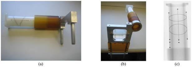
Prostate brachytherapy guided by transrectal ultrasound is a common treatment option for early stage prostate cancer. Prostate cancer accounts for 28% of cancer cases and 11% of cancer deaths in men with 217,730 estimated new cases and 32,050 estimated deaths in 2010 in the United States alone. The major current limitation is the inability to reliably localize implanted radiation seeds spatially in relation to the prostate. Multimodality approaches that incorporate X-ray for seed localization have been proposed, but they require both accurate tracking of the imaging device and segmentation of the seeds. Some use image-based radiographic fiducials to track the X-ray device, but manual intervention is needed to select proper regions of interest for segmenting both the tracking fiducial and the seeds to evaluate the segmentation results, and to correct the segmentations in the case of segmentation failure, thus requiring a significant amount of extra time in the operating room. In this paper, we preset an automatic segmentation algorithm that simultaneously segments the tracking fiducial and brachytherapy seeds, thereby minimizing the need for manual intervention. In addition, through the innovative use of image processing techniques such as mathematical morphology, Hough transforms, and RANSAC, our method can detect and separate overlapping seeds that are common in brachytherapy implant images. Our algorithm was validated on 55 phantom and 206 patient images, successfully segmenting both the fiducial and seeds with a mean seed segmentation rate of 96% and sub-millimeter accuracy.
Previous Automatic Seed Segmentation Algorithm
In a previous work we proposed a ROI-free automatic FTRAC and seed segmentation algorithm with the aims of making tools that require as little human interaction as possible. The purpose of this algorithm was to minimize the need of operator intervention to allow for automatic processing and pipelining from image acquisition to seed reconstruction and to improve upon existing FTRAC and seed segmentation algorithms for accurate three-dimensional seed localization. To evaluate the algorithm, we had 162 clinical images, and 152 images were successfully segmented (93% success rate). Overall the algorithm was successful and improved the practicality of the FTRAC, which at the time was proven better than other methods. It strengthened the prospects of using TRUS and X-ray to guide prostate brachytherapy in the OR, eliminating the current limitation of the TRUS system in being unable to localize seeds in relation to the prostate.
|
Our New Automatic Seed Segmentation Algorithm
Our proposed algorithm (Figure 2) takes a single X-ray grayscale image and outputs the equations of the lines, the 2-D image and the equations of the lines, the 2-D image coordinates of the points, and the equations of the lines, the 2-D image coordinates of the points, and the equations of the conics (note that the FTRAC has 3 parallel lines, 9 BBs, and 2 ellipses) as well as the 2-D image coordinates of the brachytheray seeds. In cases when there are overlapping seeds in a projection view, the algorithm automatically classifies them as overlapping and outputs separated image coordinates. If desired, the resulting segmentation can be overlaid on the input image for visualization (see Fig. 3). There are several assumptions made regarding the X-ray image, all of which are practical in the clinical setting: 1) the X-ray image has been corrected for geometric image distortion caused by the X-ray image intensifier, 2) the FTRAC and seeds are fully visible within the X-ray field of view (FOV), 3) the FTRAC appears to the right of the seeds without overlapping them, 4) the FTRAC is oriented upright, and 5) the TRUS probe is retracted and therefore not located in the FOV. Our algorithm was tested on Palladium-103 seeds which tend to appear small and oval in X-ray.
|
Results
The algorithm was tested on 55 phantom and 206 clinical images. The outputs of the algorithm were compared to manually corrected automatic seed segmentation of the FTRAC and the seeds. A total of 206 patient images were collected from 7 patients and the X-ray images were taken at various C-arm poses with the number of implanted seeds varying from 22 to 84 seeds. 44 images were not taken properly therefore failing to satisfy one assumption of our algorithm. Of the remaining 162 images 143 FTRAC segmentations were successful (an 88.3% success rate). For seed segmentation, 13 of the 143 images with successful FTRAC segmentations were improperly taken with the TRUS probe in the field of view. Consequently, in the remaining 130 images, our algorithm detected 7118 seeds while there were 7014 seeds. Of all the seeds detected, 6936 matched to the manually corrected segmented seeds (98.9 seed detection rate) but 2.6% of the algorithms seeds were false positives.
Conclusion
This algorithm successfully simultaneously segments the FTRATC and the seeds while effectively identifying and separating overlapped seeds. We now have a pipelined seed reconstruction system for prostate brachytherapy and minimized manual intervention caused by segmentation failures. While our algorithm is not perfect, it improvs the current workflow. At their worst, previous algorithms would require the operator to travel back and forth from C-arm to console, impending with the multiple responsibilities of operating the C-arm, specifying ROIs, Inspecting the results, and manually correcting segmentations, which can take as much as several additional minutes in the OR. Our algorithm lets the operator focus solely on the C-arm and at the algorithms best, only be limited by their speed in C-arm operation. We have presented our work for simultaneously segmenting the FTRAC and brachytherapy seeds. Through innovative use of image processing techniques, our method is able to give satisfactory results for both FTRAC and seed segmentation.
Publications
- N. Kuo, A. Deguet, D. Y. Song, E.C. Burdette, J.L. Prince, and J. Lee, "Automatic segmentation of radiographic fiducial and seeds from X-ray images in prostate brachytherapy", Medical Engineering & Physics, 34(1): 64-77, 2012.
- N. Kuo, J. Lee, A. Deguet, D. Song, E.D. Burdette, and J.L. Prince, "Automatic segmentation of seeds and fluoroscope tracking (FTRAC) fiducial in prostate brachytherapy x-ray images", Proceedings of the Society of Photo-Optical Instrumentation Engineers, 15(43), 2010.
- N. Kuo, J. Lee, C. Tempany, M. Stuber, and J.L. Prince, "MRI-Based Prostate Brachytherapy Seed Localization", Seventh IEEE International Symposium on Biomedical Imaging (ISBI 2010), Rotterdam, The Netherlands, April 14 - 17, 2010.


