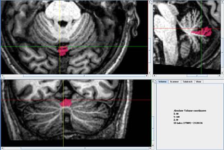
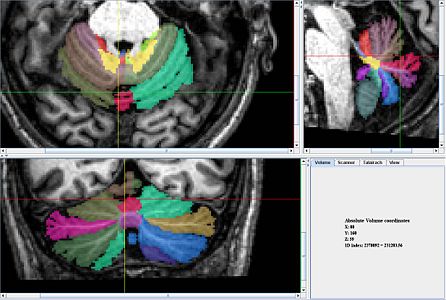
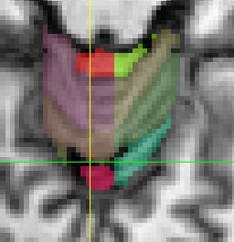
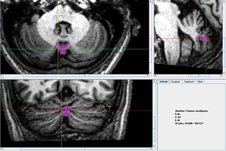
Lobules VI-VIIB vermis
Jump to navigation
Jump to search
<meta name="title" content="CHANGE ME TO THE PAGE TITLE"/>
Lobules VI-VIIB
Vermis Lobule VI
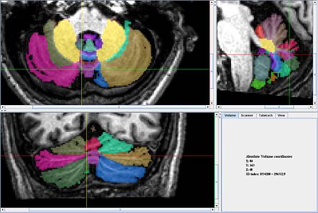
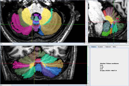
- This Lobule of the vermis is best delineated in the axial view.
- It appears first in the uppermost slices of the axial view at the midline and is often a nearly detached circle.
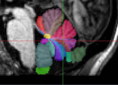
- The lower portion of this lobule of the middle vermis can best be defined in the sagittal view; at the midline the entirety of lobule VI should be vermis and its boundaries with the hemisphere should make anatomical and reasonable sense when considering the anterior portion of this vermis lobule.
- These lateral boundaries should be relatively gradual and located very near the vermis/hemisphere boundaries for the caudal lobules (shich should already be delineated.
Vermis Lobule VIIAt
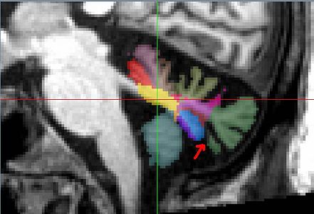
- This Lobule of the vermis is best delineated in the sagittal view.
- The entirity of lobule VIIAt should be labeled as vermis at the midline unless it appear as two detached regions.
- The lateral boundary is best defined by the increasing prominence of posterior region.
- The vermis of lobule VIIAt also ends in roughly the lateral location as the vermis of lobule VI and the caudal lobes.
Vermis Lobule VIIB
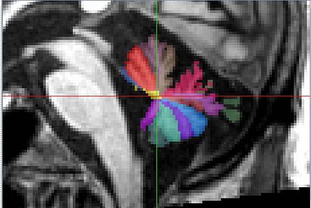
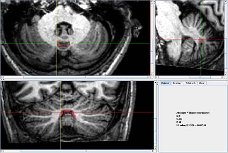
- This Lobule of the vermis is also best delineated in the sagittal view.
- It is usually much smaller than the other two lobules of Middle vermis.
- The entirity of lobule VIIB should be labeled as vermis at the midline unless it appear as two detached regions.
- The lateral boundary is best defined by the increasing prominence of posterior region.
- The vermis of lobule VIIB also ends in roughly the lateral location as the vermis of lobule VI, lobule VIIAt, and the caudal lobes.
