Difference between revisions of "Lobule VIIAf"
Jump to navigation
Jump to search
| Line 15: | Line 15: | ||
<gallery widths="500" heights="300"> | <gallery widths="500" heights="300"> | ||
image:AnonKwijibo53_unusual_Cr1.jpg|''Figure 1'':In this scan Cr1 curves around lobule VI which is very unusual. | image:AnonKwijibo53_unusual_Cr1.jpg|''Figure 1'':In this scan Cr1 curves around lobule VI which is very unusual. | ||
| − | image:AnonKwijibo56_unclearpartofCr1.jpg | + | image:AnonKwijibo56_unclearpartofCr1.jpg|''Figure 2'': The indicated portion of Cr2 seems to belong to both lobules. |
</gallery> | </gallery> | ||
Revision as of 16:43, 15 October 2009
<meta name="title" content="Lobule VIIAf (Crus I)"/>
Lobule VIIAf (Crus I)
- Location: Spans entire cerebellum except for a few slices around the midsagittal
- Description: Best to initially identify from the most lateral edges (Figure 53) and the most posterior axial slice (Figure 51)
- Notice how this lobule is not in the midsagittal slice (Figure 51)
- This lobule can also be identified in the sagittal plane as the lobule that is not present at the mid-line but becomes easily discernible roughly 15 mm from the mid-line. It is usually one branch at first that may become two or more at the lateral edges. It should dominate the lateral portion of the cerebellum.


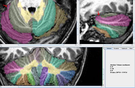
Figure 1:In this scan Cr1 curves around lobule VI which is very unusual.

Figure 2: The indicated portion of Cr2 seems to belong to both lobules.


