Protocol for Cerebellar Labeling
John A. Bogovic, Bruno Jedynak, Rachel Rigg, Annie Du, Bennett A. Landman, Jerry L. Prince, and Sarah H. Ying
Abstract
Volumetric measurements obtained from image parcellation have been instrumental in uncovering structure-function relationships. However, anatomical study of the cerebellum is a challenging task. Because of its complex structure, expert human raters have been necessary for reliable and accurate segmentation and parcellation. Such delineations are time-consuming and prohibitively expensive for large studies. Therefore, we present a three-part cerebellar parcellation system that utilizes multiple inexpert human raters that can efficiently and expediently produce results nearly on par with those of experts. This system includes a hierarchical delineation protocol, a rapid verification and evaluation process, and statistical fusion of the inexpert rater parcellations. The quality of the raters' and fused parcellations was established by examining their Dice similarity coefficient, region of interest (ROI) volumes, and the intraclass correlation coefficient of region volume. The intra-rater ICC was found to be 0.93 at the finest level of parcellation.
Online protocol document
The IACL Cerebellar Delineation Protocol can be found online, and includes:
- A guide to using MIPAV for delineation of the whole cerebellum.
- Descriptions of delineating the lobes of the cerebellum.
- Methods for parcellating the cerebellar vermis.
- A protocol for delineation of the cerebellar lobules.
Each step includes careful descriptions and screenshots of demonstrative examples.
Background
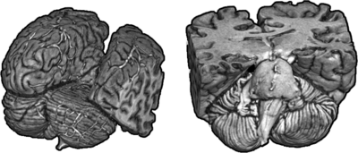
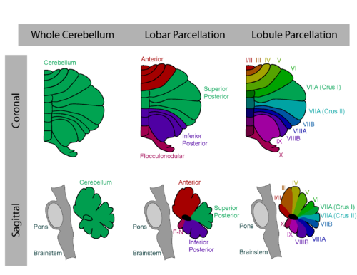
The cerebellum is a phylogenetically ancient structure connecting directly or indirectly to all elements of voluntary, postural, and ocular motor systems. It plays a central role in sensory input and voluntary motor action including limb movement, speech, and eye movement. It is located behind the brainstem and below the cerebrum (see Fig. 1). Diseases involving the cerebellum can produce symptoms such as gait and stance irregularities, tremor, eye movement problems, slurred speech, language and memory difficulties, and even personality changes. Atrophy of the cerebellum is a hallmark of many diseases that involve the cerebellum, and regional atrophy within the cerebellum is also observed. Since the cerebellum has a regular and hierarchical organization (see Fig. 2), determination of regional atrophy involving its specific lobes and lobules can be strongly indicative of specific functional deficits. The objective of this work is to define a standard manual procedure to reliably and reproducibly delineate the cerebellar lobes and lobules from MR images. This procedure can be used directly in scientific studies of cerebellar structure and function or in clinical cases. It can also be used as the basis for automatic procedures for cerebellar parcellation.
Publications
- B. A. Landman, J. Bogovic, and J. L Prince, “Efficient Anatomical Labeling by Statistical Recombination of Partially Label Datasets,” International Society for Magnetic Resonance in Medicine (ISMRM), Honolulu, Hawaii, April, 2009.
- B.A. Landman, J.A. Bogovic, and J.L. Prince, “Simultaneous truth and performance level estimation with incomplete, overcomplete, and ancillary data,” Proc SPIE 7623 Medical Imaging, Paper 7623-58, San Diego, 13-18 February, 2010.
- B.A. Landman, A.J. Asman, A.G. Scoggins, J.A. Bogovic, F.Xing, and J.L. Prince, "Robust Statistical Fusion of Image Labels," IEEE Transactions on Medical Imaging, Vol.31, No.2, pp.512-22, February, 2012. (doi) (PubMed) (PMCID 3262958).
- B.C. Jung, S.I. Choi, A.X. Du, J.L. Cuzzocreo, H.S. Ying, B.A. Landman, S.L. Perlman, R.W. Baloh, D.S. Zee, A.W. Toga, J.L. Prince, and S.H. Ying, "MRI shows a region-specific temporal pattern of neurodegeneration in SCA2," The Cerebellum, Vol.11, No.1, pp.272-279, 2012. PMID: 21850525
- B.C. Jung, S.I. Choi, A.X. Du, J.L. Cuzzocreo, Z.Z. Geng, H.S. Ying, S.L. Perlman, A.W. Toga, J.L. Prince, and S.H. Ying, "Principal Component Analysis of Cerebellar Shape on MRI Separates SCA Types 2 and 6 into Two Archetypal Modes of Degeneration," The Cerebellum, vol.11, no.4, pp.887-95, December 2012. PMID: 22258915
- J.A. Bogovic, B. Jedynak, R. Rigg, A. Du, B.A. Landman, J.L. Prince, and S.H. Ying, "Approaching expert results using a hierarchical cerebellum parcellation protocol for multiple inexpert human raters," NeuroImage, Vol.64, pp.616-629, 2013. (doi) (PubMed).


